The importance of planning and soft tissue contouring
Initial situation:
Tooth 12 had to be extracted as a consequence of severe inflammation. The crown was replaced with a Maryland bridge for the healing period. For the site of the missing tooth we produced a digital design for the further steps, by mirroring the lateral incisor on the opposite side.
On the basis of the Maryland bridge and the preliminary design, a surgical guide was made for the precise placement of a Straumann Bone Level NC implant. Using it, the implant was placed in an ideal position from both a biomechanical and a prosthetic point of view. After placement the temporary bridge was re-bonded for the healing period.
After uncovering the implant, contouring of the gingiva followed. For this a temporary crown was made in the practice with the help of the preliminary design and a 3D printed model.
The shaping of the temporary crown decisively determines the healing of the soft tissues, so it is critical for the quality of the definitive crown as well.
We shaped the subgingival parts, while leaving room for the papillae to regenerate. During healing we managed to establish favourable gingival conditions.
To reproduce the emergence profile, a custom impression component was made, with the help of which we could model the situation precisely on the stone model.
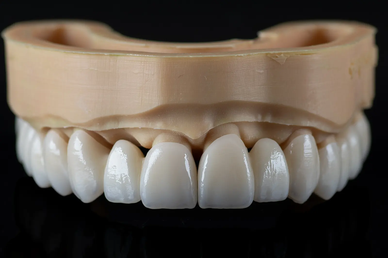
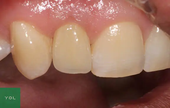
The model was then scanned and we designed a zirconia-ceramic crown that received ceramic veneering only on the labial surface. In this way we could produce a durable crown with protected incisal edges that is nevertheless aesthetic. The subgingival part is strictly only mechanically polished, and the contour of the crown corresponds to the contoured gingiva.
Images taken after delivery:
Surgical guide design and prosthetic work: dr. Molnár Bertold
Implant placement: dr. Németh Bálint